Telomere Dysfunction in Human Epithelial Tumorigenesis: Early, Widespread, and Insufficient
A thesis reconstruction.
About this reconstruction
This is the thesis that the existing data already supports, assembled from four published papers and nothing else. No experiment here is hypothetical.
The work spans a PhD program at Indiana University School of Medicine, 2009-2013, in the Gilley lab. The degree was not completed. What follows is an argument that could have been defended on the data as it stood, reorganized around a claim the individual papers state but never foreground.
Contribution levels differ across the four papers, and that matters for what a thesis could legitimately claim:
| Paper | Position | Contribution | Thesis role |
|---|---|---|---|
| Tu et al. 2015, Mol Carcinogenesis | Co-first author | Most of the work | Core data chapter |
| Tanaka et al. 2012, PNAS | Fourth of seven | Substantial | Methods + breast chapter, carved to own contribution |
| Kannan, Huda, Tu et al. 2013, Stem Cell Reports | Third of twelve | Substantial | Collaborative chapter, framed as such |
| Williamson et al. 2012, EMBO Mol Med | Eighth of thirteen | Minor | Not included |
The PARP/ATM paper is left out deliberately. It is good work on a different question, and a thesis is stronger for excluding a chapter that does not serve its argument.
One honest caveat throughout: the synthesis in Chapter 6 is a reinterpretation, not a new result. Its strength is that it requires no data beyond what exists, and its weakness is that the decisive experiment distinguishing its model from the alternatives was never run.
Abstract
Telomere dysfunction is not the rate-limiting step in human epithelial carcinogenesis. Measured directly, in human tissue, with a single assay, across three systems, telomere fusion burden fails to distinguish benign from malignant at every point where the standard model predicts it should.
The breakage-fusion-bridge model holds that critically short telomeres drive end-to-end chromosome fusion, that fusion drives genomic instability through successive breakage cycles, and that this instability drives malignant progression. The model is mechanistically sound and well supported in engineered systems. It has rarely been tested directly in human tissue, because telomere fusions themselves were hard to measure there.
This thesis applies TAR-fusion PCR, a multiplex assay that amplifies and sequences chromosome end-to-end fusion junctions from solid tissue DNA, across a deliberately chosen gradient, and reports three results that do not fit.
First, in breast, telomere fusions are absent from normal tissue and present in roughly 40% of ductal carcinoma in situ. They are also present in roughly 40% of invasive ductal carcinoma. Fusion burden marks the onset of neoplasia and then does not change across the transition that defines the disease.
Second, in normal breast, the luminal progenitor compartment carries a mean telomere length of 3.4 kb and a telomere-specific DNA damage response in over 95% of cells, roughly ten dysfunction-induced foci per nucleus, and no detectable fusions, by either PCR or metaphase analysis. A quarter to a half of these cells remain clonogenic. The compartment with the most dysfunctional telomeres in the normal breast is held indefinitely at the threshold without crossing it.
Third, in prostate, benign prostatic hyperplasia is fusion-positive in 65% of samples, against 55% for the established precursor lesion PIN and 62% for prostate cancer. BPH also carries anaphase bridges, aneuploidy and telomerase activity. A lesion with essentially no malignant potential is indistinguishable from carcinoma on every measure of telomere-driven instability applied here.
Taken together: telomere dysfunction is early, widespread, and insufficient. It occurs in normal tissue, in benign disease, in precursor lesions and in carcinoma, at frequencies that do not order those states. The informative variable is therefore not whether telomere dysfunction occurs, but what restrains its consequences. Three candidate restraints are identified in the data presented here: compartment-specific telomerase salvage, checkpoint-enforced cell cycle exit, and the clonal versus stochastic character of the fusion events themselves. A model is proposed in which these gates, rather than telomere dysfunction itself, determine which cells with dysfunctional telomeres contribute to tumorigenesis.
Chapter 1: Introduction
The gap this thesis addresses is evidentiary, not conceptual: the breakage-fusion-bridge model of telomere-driven carcinogenesis rests largely on engineered systems and on indirect markers in human tissue.
What telomeres do
Telomeres are tandem TTAGGG repeats with associated proteins that disguise the chromosome terminus from recognition as a double-strand break. Without that disguise, ends are joined to other ends. Telomeric DNA is synthesized by telomerase, which is active in germline and primitive compartments and generally absent at maintenance levels from differentiated somatic cells.
The standard model
Critically short telomeres trigger senescence or apoptosis. Where those responses are disabled, ends fuse; the resulting dicentric chromosomes break in anaphase; the broken ends fuse again. Successive breakage-fusion-bridge cycles generate the non-reciprocal translocations, amplifications and aneuploidy characteristic of carcinoma. Telomerase reactivation then stabilizes the genome, freezing the rearrangements already acquired.
The model is attractive because it explains the right things: why most human cancers are carcinomas of high-turnover epithelia, why instability appears early, and why telomerase is reactivated in the large majority of tumors.
What the evidence actually was
Support came from three sources, none of them a direct measurement of fusion in human tissue.
- Mouse genetics. Telomerase-null mice on a p53-mutant background develop breast and colon carcinomas, establishing that telomere dysfunction is sufficient to drive epithelial tumorigenesis in that system.
- Cell culture. Late-passage human mammary epithelial cells that escape a stress-associated senescence barrier acquire genomic alterations including telomere fusions.
- Indirect markers in human tissue. Shortened telomeres by FISH in luminal cells and DCIS, and anaphase bridges in early lesions.
The third category is the weak link. Short telomeres identify cells at risk of dysfunction; anaphase bridges are consistent with dysfunction but not specific to it. Neither establishes that fusion has occurred. Direct evidence of telomere dysfunction in human solid tumors had been reported only in chronic lymphocytic leukemia, where circulating cells make the DNA tractable.
The question
If telomere fusions can be measured directly in human solid tissue, three questions follow immediately, and this thesis asks them in order:
- At what stage of carcinogenesis do fusions first appear?
- Do normal cells with critically short telomeres carry fusions?
- Does fusion burden distinguish lesions by malignant potential?
The standard model answers the first two with conditions attached. Fusions appear once checkpoints fail, which in a progressing lesion means early and then increasingly; and normal cells with short telomeres should arrest rather than fuse, because their checkpoints are intact. Only the third answer is unconditional: fusion burden should rise with malignancy. The chapters that follow confirm the arrest prediction, find the first answer to be “early, and then flat,” and contradict the third outright. The pattern that emerges is that the model is right wherever it attaches a condition and wrong wherever it does not, which is the subject of Chapter 6.
Chapter 2: TAR-fusion PCR
TAR-fusion PCR amplifies chromosome end-to-end fusion junctions directly from solid tissue DNA, converting telomere dysfunction from an inference into a sequenced observation.
Design
The assay uses multiplex primer sets annealing within telomere-associated repeat 1 (TAR1) regions, which lie within roughly 2 kb of the telomere variant repeat regions at chromosome ends and share partial homology with one another. Two multiplex sets are used to increase coverage of possible fusion combinations while limiting reaction number. Two additional primers anneal within distal Xp and 17p, outside known TAR1 regions.
Coverage is the assay’s principal limitation and is stated plainly: the primer sets address approximately 14% of theoretically possible chromosome end-to-end fusion combinations. The constraint is not methodological but informational: sequence for the majority of chromosome termini was absent from genome assemblies.
This limitation matters in one direction only. Every frequency reported in this thesis is a floor, not an estimate. A sample scored fusion-negative may carry fusions at uncovered ends; a sample scored positive carries at least one real, sequence-confirmed fusion.
Validation
The assay was developed against the BJ foreskin fibroblast HPV16 E6/E7 telomere crisis model, which accumulates fusions with increasing population doublings. Three features establish that it amplifies genuine fusions rather than PCR artifact.
| Population | Mean TRF | Dicentrics per metaphase |
|---|---|---|
| BJ, 50 PD | 7.6 kb | 0.20 |
| BJ E6/E7, 87 PD | 5.9 kb | 1.15 |
| BJ E6/E7, 96 PD | 5.2 kb | 2.95 |
First, product accumulation tracks the independently measured dicentric frequency across this series. Second, junction sequence analysis confirms fusion architecture directly. Third, primary BJ fibroblasts and the large majority of normal human genomic DNA give no signal under the optimized conditions.
What the junctions look like
Sequencing showed that 78.6% of products contained shortened telomeric DNA, roughly 60 to 700 bp, adjacent to subtelomeric DNA at the junction. The majority of telomeric tracts terminated in two or three G residues.
Two points follow. Telomeric DNA is substantially shortened before fusion, consistent with attrition as the trigger. And the junction’s composition reports on the pathway that joined the ends, not on the structure of the end before it was joined, a distinction that becomes important in Chapter 3.
One qualitative difference from the previously reported fusion PCR method: TAR-fusion PCR detects telomere-to-telomere junctions, not only telomere-to-subtelomere.
Scope of this chapter’s contribution
Assay design and primary development were led by others in the laboratory. The work presented here as the candidate’s own concerns junction sequence analysis and application of the validated assay to tissue, which is where Chapter 3 begins.
Chapter 3: Breast carcinoma
Fusion burden marks the onset of neoplasia in the breast and then stops being informative: ductal carcinoma in situ and invasive ductal carcinoma are fusion-positive at the same frequency.
Result
| Tissue | n | Fusion-positive |
|---|---|---|
| Normal breast, healthy volunteers | 23 | 0% |
| Ductal carcinoma in situ, grade 3 | 25 | ~40% |
| Invasive ductal carcinoma | 23 | ~40% |
The normal-tissue result is as important as the lesion results. Zero of twenty-three establishes that the assay does not report background, and it sets the baseline against which Chapter 4 reads.
The early finding, and the flat one
The early appearance of fusions in DCIS is the result the standard model predicts, and it was the headline. Telomere dysfunction is present before invasion, alongside previously reported telomere shortening, telomerase activation and other genomic aberrations in DCIS.
The flat finding was not foregrounded, and it is the more informative of the two. Between in situ and invasive disease, the transition that defines clinical breast cancer, fusion burden does not move. Whatever distinguishes a lesion that stays within the duct from one that does not, it is not how much telomere fusion the lesion carries.
One reading preserves the standard model: telomere dysfunction may be actively limited at later stages by telomerase reactivation, preventing a genomic catastrophe incompatible with tumor cell viability. Under that reading, flat burden reflects a brake applied after the damage is done. This reading is plausible and is not excluded by these data. It is also, notably, a gate, which is the thesis of Chapter 6.
Junction architecture changes even where burden does not
Sequence analysis of fusion junctions revealed two features that do differ between stages, and they are the chapter’s strongest result.
Microhomology at the junction was present in the crisis model and in invasive tissue, but not in DCIS grade 3. This suggests that a portion of fusions in invasive tissue arise through end-joining pathways distinct from those operating in DCIS, potentially DNA-PKcs-independent non-homologous end joining or microhomology-mediated end joining.
Small regions of LTR and non-LTR retrotransposon elements, derived from interstitial chromosomal regions, were found within fusion junctions in invasive tissue. These more complex junctions are consistent with products of extensive breakage-fusion-bridge cycling. Comparable retrotransposon elements occur at Drosophila chromosome ends and have been reported to integrate at dysfunctional mammalian telomeres, raising the possibility of a conserved integration pathway at uncapped ends.
The pattern across these two observations is the chapter’s contribution to the thesis: the quantity of telomere fusion is flat across progression while the quality of fusion changes. What differs between in situ and invasive disease is not how often ends fuse but how they are processed when they do.
Limitations
Bulk tumor tissue was assayed, with tumor cell content ranging from 50%. Fusions arising in stromal cells cannot be excluded, and the cellular subpopulation carrying the fusions was not resolved. Fusion frequency was not related to tumor grade, receptor status or outcome. Both limitations are addressed in Chapter 7.
Chapter 4: The luminal progenitor
The compartment with the shortest telomeres and the heaviest DNA damage burden in the normal human breast carries no telomere fusions at all. This is the thesis’s central negative result.
Approach
Four populations were sorted to over 95% purity from reduction mammoplasty tissue across 37 donors of varying age, and analyzed without culture: basal cells (BC), luminal progenitors (LP), mature luminal cells (LC), and stroma (SC). Telomere length was measured four independent ways, by TRF Southern, qPCR, Flow-FISH on single cells, and Q-FISH on metaphases, which matters, because the headline result is a length measurement.
Luminal progenitors have critically short telomeres, independent of age
| Population | Mean telomere length | Age dependence |
|---|---|---|
| Luminal progenitor | 3.4 kb (Q-FISH) | None (R² = 0.002, P = 0.9) |
| Basal cell | 6.3 kb (Q-FISH); 6 to 8 kb by Southern | None |
| Mature luminal | ~9 kb by Southern | Declines with age (R² = 0.5, P < 0.01) |
| Stroma | Longest of the four | None |
Southern analysis implies that some chromosomes within the LP population likely carry terminal restriction fragments under 3 kb, a reading the Q-FISH data independently support. Lengths in that range have been associated with telomere dysfunction and cell death in the absence of telomerase. The LP value is age-independent, which means this is not an attrition phenotype accumulated over a lifetime. It is a steady-state property of the compartment.
One methodological note with consequences: the age-related change in LC telomere length is invisible in unseparated breast tissue, masked by the predominance of other cell types. Compartment-level biology in this tissue is simply not accessible to bulk measurement.
They carry a chronic, telomere-specific DNA damage response
NBS1, RAD50, γH2AX and 53BP1 were essentially undetectable in basal cell nuclei and strongly present in every luminal progenitor examined, consistently colocalized with TRF2, at roughly ten telomere dysfunction-induced foci per nucleus. Over 95% of LPs show this; under 5% of basal cells do. MRE11 colocalized with TRF2 in some basal nuclei but significantly less than in LPs (P < 0.00001).
The transcriptome agrees. Of 50 telomere-associated genes differentially expressed between LP and BC, thirteen were elevated in LPs, including MRE11, RAD50, ATM, ATR and BLM. RAP1 and DNA-PK were higher in basal cells.
For scale: senescence has been reported to follow accumulation of five or more such foci per cell. Luminal progenitors carry roughly twice that, as a normal condition, throughout adult life.
And no fusions
Neither TAR-fusion PCR on freshly isolated LPs and BCs from four donors of varying age, nor examination of metaphases prepared for Q-FISH, revealed any evidence of telomere fusion.
This is not, on its own, a refutation of the standard model. That model holds that ends fuse where senescence and apoptosis have been disabled, and luminal progenitors are checkpoint-intact: the elevated P14, P53, PML, ID2 and E2F1 reported below are the response it expects. Arrest, not fusion, is the predicted outcome, and the source paper reads it the same way, concluding that these cells “appear to be protected from such events, at least to some extent,” by mechanisms that push them out of cycle.
What the result does establish is magnitude. A normal compartment sustains telomeres at 3.4 kb and twice the damage burden associated with senescence onset, indefinitely, without producing the fusions that a benign lesion produces readily. That sets the floor against which Chapters 3 and 5 are read, and it locates the interesting variable in the restraint rather than in the dysfunction.
Two restraints are visible in the data
Telomerase salvage. Telomerase activity was elevated only in LPs among the four populations. hTERT transcript was consistently and selectively elevated, with roughly three hTERT-TRF2 colocalized foci per nucleus in 80% of LPs. Critically, this activity is highest in younger women and declines with age, the only element of LP telomere biology that moves with age at all.
Checkpoint-enforced exit. LPs showed higher lysosomal content and elevated P14, P53, PML, ID2 and E2F1, with decreased TBX2 and TBX3. The interpretation offered was that cells reaching critically short telomeres are removed from cycle, which would explain both the absence of fusions and why higher LP clonogenic frequencies have never been achievable in culture.
The paradox that remains
Between a quarter and nearly half of cells with the LP phenotype form colonies in vitro, while over 95% carry roughly ten dysfunction-induced foci. A substantial fraction of these cells are proliferating with a telomere damage response that should have arrested them.
The compartment is therefore not simply protected. It is held at the threshold, damaged and cycling and not fusing, by something active. Identifying that restraint is the single most valuable unfinished experiment arising from this work, and Chapter 7 specifies it.
Why this compartment matters beyond telomere biology
Luminal progenitors are the proposed cell of origin for BRCA1-associated breast cancer, with malignant cells carrying a transcriptional profile resembling normal LPs despite a basal phenotype. The compartment that gives rise to those tumors is the compartment carrying the most dysfunctional telomeres in the normal breast. Whether that coincidence is causal is unresolved and, as Chapter 7 argues, directly testable.
Limitations
The central negative in this chapter is a limit of detection, not a demonstrated absence. TAR-fusion PCR was run on four donors, at 14.2% coverage of possible fusion combinations, using 50 ng of template per reaction. That is roughly 7,500 diploid genome equivalents, so the assay reports fusions that are abundant in a sample, which in practice means clonal or occurring at high per-cell frequency. A private fusion in a single luminal progenitor would be one template copy among thousands and would not be expected to amplify.
The template load is at least matched across this thesis. Chapters 3 and 5 used the same 50 ng per reaction, so the zero reported here and the 65% reported for benign prostatic hyperplasia are not an artifact of unequal input. The metaphase analysis adds less than it appears to, because those metaphases were prepared for Q-FISH rather than scored for fusion at a stated count.
The claim this chapter supports is therefore that no abundant or clonal fusion is detectable in luminal progenitors, not that luminal progenitors never fuse.
Scope of this chapter’s contribution
This work was a collaboration with the Terry Fox Laboratory, which supplied cell sorting and Flow-FISH capability. The candidate’s contribution centered on the telomere dysfunction and fusion analyses.
Chapter 5: Prostatic lesions
A lesion with essentially no malignant potential carries telomere fusions at the highest frequency of any tissue examined in this thesis, along with anaphase bridges, aneuploidy and telomerase activity.
Result
| Lesion | Fusion-positive | Malignant potential |
|---|---|---|
| Benign prostatic hyperplasia | 65% | Negligible |
| High-grade prostatic intraepithelial neoplasia | 55% | Established precursor |
| Prostate cancer | 62% | Malignant |
| Tumor-adjacent, histologically normal | Generally undetectable | n/a |
Anaphase bridges and aneuploidy followed the same pattern, present across all three lesion types and generally absent from tumor-adjacent normal tissue. Telomerase was detectable in BPH and PIN, with higher average levels in PCa. Given the assay’s 14% coverage ceiling, these frequencies are floors, which makes the convergence more striking rather than less.
Why this is the decisive result
BPH is the control the standard model did not anticipate. It originates in basal epithelial cells, it is extremely common, and the prevailing consensus is that it does not progress to prostate cancer. It is therefore a lesion in which proliferation, telomere dysfunction, chromosomal instability and telomerase activation coexist indefinitely without malignant outcome.
The finding in PIN supports the conventional interpretation: fusions, bridges, aneuploidy and telomerase in an established precursor are consistent with its precursor status. The finding in BPH removes that interpretation’s force. The same marker set, at equal or higher frequency, in a lesion that goes nowhere, means the markers do not identify what is going somewhere.
Stated as the original paper did: genomic instability and telomerase activation alone are not sufficient for malignant transformation, and other barriers must exist to prevent the vast majority of BPH lesions from progressing.
That sentence is this thesis’s argument, and this chapter is where the data compel it.
The tumor-adjacent anomaly
Histologically normal tissue adjacent to prostate carcinoma showed no detectable telomere fusions, but did show relatively high telomerase activity correlating closely with the matched tumor (R² = 0.72 across 15 pairs). The internal control is strong: hTERT transcript and telomerase activity correlate at R² = 0.98, so the telomerase measurement is sound.
Two explanations were offered and neither was resolved. Infiltrating lymphocytes express moderate telomerase levels. Alternatively, this is field cancerization, well documented in prostate, with telomerase activation in tumor-adjacent normal tissue reported in head and neck and in breast.
The second explanation has a consequence worth stating explicitly, because it was not. If the telomerase is epithelial, then telomerase activation extends across a histologically normal field in the absence of detectable telomere fusion, which inverts the assumed order of events. The standard sequence runs shortening, then crisis, then fusion, then telomerase rescue. These data would instead place telomerase activation first, across a field, with fusion arising later in a subset. Distinguishing the two requires only CD45 depletion before the telomerase assay, or hTERT immunohistochemistry to localize the signal.
Limitations
Measurements are at whole-tissue level and do not reflect per-cell comparison, since the proportion of target epithelial cells to unaffected cells varies between samples and between lesion types. This is the same bulk-tissue constraint as Chapter 3, and the same remedy applies.
Chapter 6: Synthesis
Read across all three systems, telomere fusion burden has no discriminating power. It is zero where telomeres are shortest, highest where malignant potential is lowest, and flat across the transition that defines malignancy.
The comparison
The three preceding chapters were conducted as separate studies. Their value is greatest when read as one dataset, because they share an assay, a laboratory and a scoring standard, a comparison that is normally unavailable across tissues, since cross-study fusion frequencies are not commensurable.
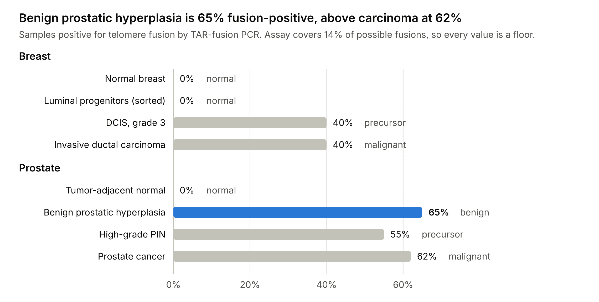
The series is not monotonic in malignant potential at any point. The two highest values belong to a benign lesion and a carcinoma; the two zeros belong to normal tissue and to the compartment with the shortest telomeres measured anywhere in this work.
What the comparison rules out
Three readings of this dataset do not survive scrutiny. The standard model is not among the casualties. It survives all three, but what survives is a model whose predictive work is done entirely by the conditions it attaches, and not by telomere dysfunction itself.
That fusion burden measures progression. It does not. BPH at 65% exceeds PCa at 62%, and DCIS equals IDC. Any model in which accumulated fusion drives advancement predicts a monotonic series, and the series is not monotonic.
That telomere length alone orders tissues by fusion burden. It does not. The shortest telomeres measured anywhere in this work, luminal progenitors at 3.4 kb with ten damage foci per nucleus and retained clonogenicity, sit in the compartment with no detectable fusions, while a benign prostatic lesion with no such extremity of length is fusion-positive in two thirds of samples. This is not a refutation of the standard model, which requires checkpoint loss before ends fuse and so predicts exactly this in normal tissue. It does rule out any simpler reading in which shortness itself is what the fusion data track.
That telomerase reactivation is a late event marking escape. It is not, at least not uniformly. Telomerase is present in normal luminal progenitors, in BPH, in PIN, and across histologically normal tissue adjacent to prostate carcinoma where no fusions are detectable at all.
What the comparison puts in their place
If telomere dysfunction is common to normal tissue, benign disease, precursor lesions and carcinoma alike, then its presence cannot be what separates them. Something downstream must. This thesis identifies three candidate restraints, each visible in the data presented, and proposes that these gates rather than dysfunction itself determine outcome.
Gate 1, telomerase salvage. Compartment-specific telomerase permits survival at telomere lengths that would otherwise be lethal. Luminal progenitors sustain 3.4 kb telomeres indefinitely and show selectively elevated hTERT with telomere-localized foci in 80% of cells. This activity declines with age, making it the only age-variable element of LP telomere biology and therefore a candidate clock.
Gate 2, checkpoint-enforced exit. Cells reaching critically short telomeres leave the cycle before fusion can occur. LPs carry elevated P14, P53, PML, ID2 and E2F1 and increased lysosomal content. This gate predicts that the fusion-free state of normal LPs is actively maintained and should fail on p53 disruption, consistent with the greater than 90% TP53 mutation rate in BRCA1-associated breast cancer, whose cell of origin is this compartment.
Gate 3, clonality. This gate is a hypothesis rather than an observation, and it is the one this thesis cannot close. The assay reports whether fusions are abundant in a sample, not whether they are the same fusion, and those are different things. A lesion in which many cells each sustain a different fusion, and a lesion descended from one founder that sustained a single fusion, can both score positive at similar frequencies while meaning opposite things. Abundance is what Chapter 5 measured. Diversity is what it could not. The proposal is that BPH is the first case and carcinoma the second, that junction diversity, not fusion burden, marks the transition, and that burden looks uninformative across these tissues precisely because it sums over both.
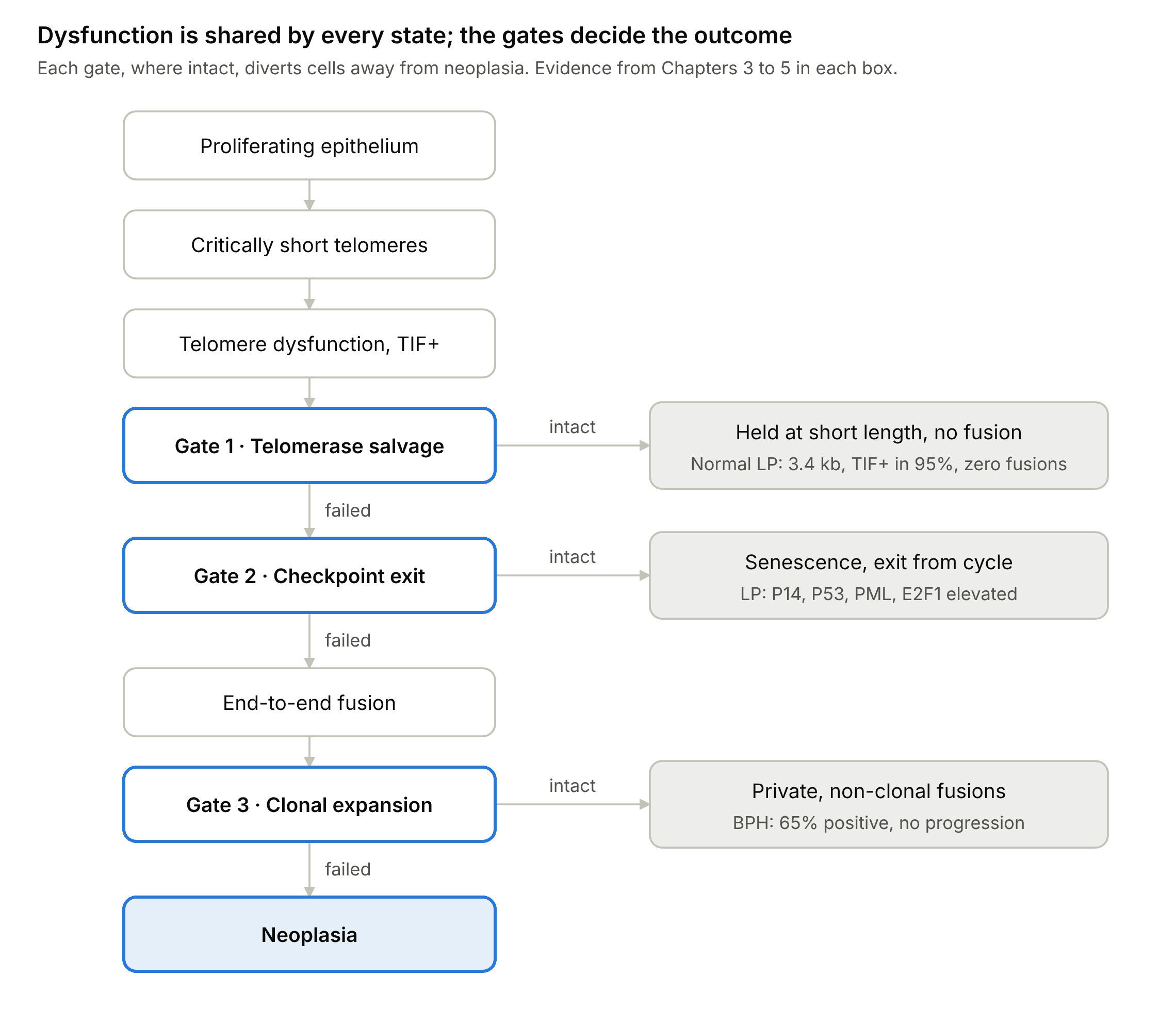
The model’s claim is that every state examined in this thesis enters at the top and that tissues differ only in which gates hold. Normal luminal progenitors are diverted at Gates 1 and 2. BPH passes both, fuses, and is diverted at Gate 3. Carcinoma passes all three.
The thesis statement
Telomere dysfunction is a permissive background condition of proliferating human epithelium rather than a rate-limiting driver of carcinogenesis. It arises early, occurs widely in tissue that never becomes malignant, and does not increase with malignant progression. What distinguishes tissues and lesions is not whether their telomeres fail but which restraints on the consequences of that failure remain intact.
What would falsify this
The model earns its place only if it can be wrong, and it can be, in three specific ways.
- If fusion junctions in BPH prove clonal rather than private, Gate 3 fails and the burden-based model is rehabilitated.
- If p53 disruption in sorted luminal progenitors does not produce fusions, Gate 2 is not the restraint holding that compartment.
- If telomerase inhibition in luminal progenitors does not shorten telomeres further or precipitate fusion, Gate 1 is not performing salvage.
Each is a discrete experiment on material the laboratory possessed. Chapter 7 specifies them.
Chapter 7: Future directions
Every experiment below uses material and methods the laboratory already had. They are ordered by what they would have cost to run, not by what they would have been worth.
1. Clonality of fusion junctions in matched prostate lesions
The decisive test of Gate 3, and the one experiment that would convert Chapter 5’s negative into a positive claim.
Take multiple spatially separated regions from the same lesion. Amplify and sequence fusion junctions from each. Ask whether junctions are shared across regions or private to them.
Prediction: BPH yields many distinct junction sequences, independent end-joining events in scattered cells, with little or no overlap between regions. PIN and PCa yield the same junction across regions, inherited from a founder that survived and expanded.
Samples, assay and sequencing protocol were all in hand. Outcome either way is publishable: a positive result establishes clonality as a candidate biomarker; a negative result rehabilitates the burden model and is equally worth knowing.
2. Resolving the tumor-adjacent telomerase signal
The R² = 0.72 correlation between tumor and matched adjacent normal hTERT is the strangest unexplained observation in this body of work.
CD45 depletion before the telomerase assay, or hTERT immunohistochemistry, distinguishes infiltrating lymphocytes from an epithelial field effect. If epithelial, telomerase activation precedes detectable telomere fusion across a histologically normal field, which reverses the canonical event order.
Modest cost, real conceptual payoff, and a defined risk: the answer may simply be lymphocytes.
3. What gates the luminal progenitor
Three arms, testing the two restraints visible in Chapter 4 directly.
- Age. Sort LPs from older donors, where telomerase has declined, and test for fusions. This is a prediction internal to the existing dataset and requires no perturbation at all, only tissue already being collected.
- p53. Knock down p53 in sorted LPs in short-term culture, then run TAR-fusion PCR. If fusions emerge, Gate 2 is confirmed and the link to TP53 mutation in BRCA1 tumors becomes mechanistic.
- Telomerase. Deplete hTERT in sorted LPs and ask whether telomeres shorten further and fusions appear. Tests Gate 1.
Primary LP culture is difficult and sample access depended on an external collaboration, which makes this the highest-value and highest-risk item on the list.
4. Comparative junction architecture across tissues
Chapter 3 found microhomology at invasive breast junctions but not DCIS, and retrotransposon elements at invasive junctions only. Whether that shift in end-joining mechanism is breast-specific or general is unknown and answerable by sequencing prostate junctions and comparing spectra.
Lowest risk of anything here: samples collected, methods established, a result guaranteed. Also the lowest ceiling.
5. The luminal progenitor in BRCA1 mutation carriers
The most ambitious extension, and the one that connects this work to the question of why BRCA1 predisposes so specifically to breast and ovarian cancer.
Apply the Chapter 4 panel to histologically normal breast tissue from BRCA1 carriers against non-carriers, sorted by compartment, across the age range rather than at a single age. The non-carrier baseline already exists in the 37-donor dataset.
The primary endpoint should be fusion, because it is binary against a true zero and therefore robust at the small sample sizes carrier tissue permits. The essential control is LOH genotyping of the sorted cells: without it, haploinsufficiency cannot be distinguished from rare second-hit clones, and no result is interpretable.
Most likely outcome is no difference, which would itself be the first compartment-resolved test of BRCA1 haploinsufficiency in human breast, and would establish that the LP’s vulnerability is lineage-intrinsic rather than mutation-derived. That framing has to be chosen before the experiment runs, not after.
Source publications
All data in this document come from these four papers. No figures or frequencies have been estimated, extrapolated or taken from elsewhere.
Chapter 2 and Chapter 3. Tanaka H, Abe S, Huda N, Tu L, Beam MJ, Grimes B, Gilley D. Telomere fusions in early human breast carcinoma. Proceedings of the National Academy of Sciences 2012; 109(35): 14098-14103. doi:10.1073/pnas.1120062109 · free full text
Chapter 4. Kannan N, Huda N, Tu L, Droumeva R, Aubert G, Chavez E, Brinkman RR, Lansdorp P, Emerman J, Abe S, Eaves C, Gilley D. The luminal progenitor compartment of the normal human mammary gland constitutes a unique site of telomere dysfunction. Stem Cell Reports 2013; 1(1): 28-37. doi:10.1016/j.stemcr.2013.04.003 · free full text
Chapter 5. Tu L, Huda N, Grimes BR, Slee RB, Bates AM, Cheng L, Gilley D. Widespread telomere instability in prostatic lesions. Molecular Carcinogenesis 2015. doi:10.1002/mc.22326
Not included. Williamson CT, Kubota E, Hamill JD, Klimowicz A, Ye R, Muzik H, Dean M, Tu L, Gilley D, Magliocco AM, McKay BC, Bebb DG, Lees-Miller SP. Enhanced cytotoxicity of PARP inhibition in mantle cell lymphoma harbouring mutations in both ATM and p53. EMBO Molecular Medicine 2012. doi:10.1002/emmm.201200229 · free full text